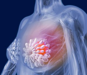
Breast cancer can happen in both men and women. But it is the most prevalent type of cancers that is diagnosed among women. It can be detected as a lump in breast tissue that is different from the rest. Other signs may include: tissue thickening/ one breast becoming larger or smaller/ nipple changing position / pain in part of breast or armpit and etc.
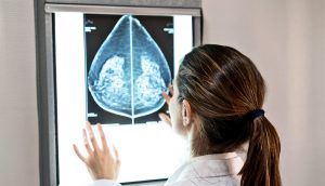
Biopsy of the affected area of breasts is used to diagnose most types of breast cancers. Moreover, there are other types of breast cancer that need to be examined with specialized laboratory methods. Since 1990s, several methods have been used for the early detection of breast cancer, such as mammography and MRI. Mammography is the standard method for early detection of breast cancer, but it has one main limitation: that is, it may produce a large number of false positives. Here we will take a look at what Mammography is:
“ Mammography is breast cancer screening using low-dose x-rays. Medical organizations suggest having mammograms every year beginning at age 40. Before women can experience any signs of cancer, mammography can diagnose it. This method can also help women to find breast diseases such as a lump, pain or nipple discharge.”
In contrast, respective standard screening techniques are used for early detection of breast cancer such as Thermography. Thermal infrared images are used to detect lesions in breasts. These Thermograms detect the temperature of the different regions of the breast. Regions with warmer temperature are more likely to contain tumors than a normal tissue. This technique has several substantial benefits:
- It is less expensive than the mammography and magnetic resonance imaging (MRI)
- It is non-contact, non-invasive
- It is non-radiative
- It is a safe diagnostic procedure, in which patients feel no pain
- This method can be used from far way
- It is possible to simultaneously monitor a large area of the population
- Interpretation of thermogram’s colors is easy and fast.
- This method only records natural radiation from the surface of the skin and there is no trace of harmful rays, so is suitable for long-term and repeating use.
- It is a fast way to monitor and observe the dynamic changes in temperature.
- Early detection up to 10 years before the cancer turns into a full-blown size
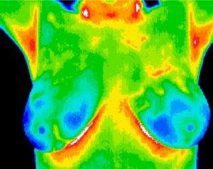
Since the normal temperature range of human body is between 36.5∘C to 37.5∘C, by using this technique, thermal infrared cameras are able to capture the variation of temperature between normal and abnormal breast tissues.
AI application for breast cancer Thermography
According to several studies, by using highly well-aimed and accurate soft-wares, breast cancer can be detected and treated at early stages of the disease. Several applications have been found in the medical profession for thermal imaging. In recent years, due to advancements in image processing techniques, Thermography has been able to achieve more than other methods such as Mammography. Thermography soft-wares (CAD) serves the need of physicians for an Al-guided support for breast cancer Thermography, which is an decisive factor in validating what they speculate, detecting what they potentially did not observe throughout the process and taking notice of highly dubious studies and researches that need to be brought to more attention.