Author: content

content
January 9, 2021
Machine learning and infrared thermal imaging in breast cancer detection
January 9, 2021

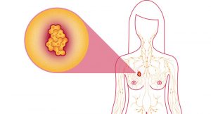
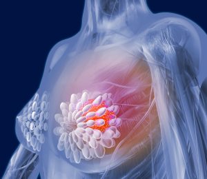
Cancer is the most important public health issue in the world and breast cancer is the most commonly cancer type detected in American women. Breast cancer is the second type more common cancers in the world and is more widespread among women.

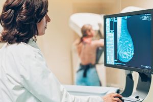
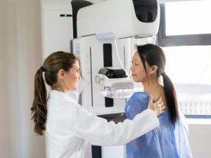
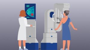
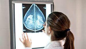
Over the past 20 years, many applications have been found in the medical profession for early detection of breast cancer such as mammography which is often used for breast cancer diagnosis.

However, the potential side effects of using mammography and the rate of false positives discourages patients and physicians to use this method. Moreover, mammography has some limitations such as the difficulty of detecting tumors in young patients or cancers without masses, such as Paget’s carcinoma. Since the young breast is mainly composed of glandular tissue, which makes it denser, this high density of the breast interferes in the identification of masses and micro-calcifications by X-rays.

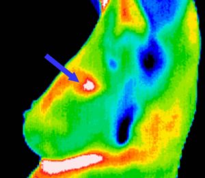
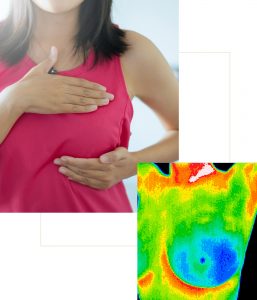
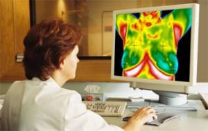
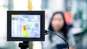
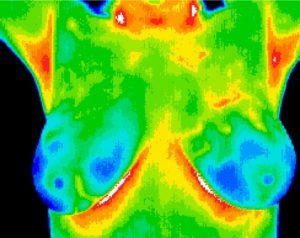
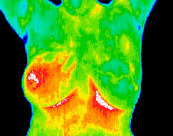
Infrared thermography (IRT) is another technique that has been used in combination with other screening techniques in the aid of breast cancer detection. This method is:

  • A low-cost technique that does not involve harmful radiation to humans
  • It does not involve invasive procedures
  • It is based on the principle of measuring the infrared radiation emitted by an object or surface through an infrared camera to determine its temperature
  • It may provide better results for breast cancer detection in young women, who usually have denser breasts, when compared to mammography

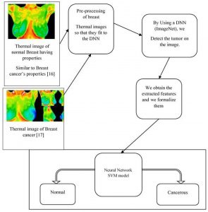
The approach proposed in this article: Gonçalves, C.B; Leles, A.C.Q.; Oliveira, L.E.; Guimaraes, G.; Cunha, J.R.; Fernandes, H. Machine Learning and Infrared Thermography for Breast Cancer Detection. Proceedings 2019, 27, 45 involves the acquisition of infrared images of healthy patients and patients with cancer followed by the extraction of features to describe these images.

Database

The database used contains data from 70 patients with or without breast tumor. These images were collected in a five-month period. The images were captured with a mid-wave infrared camera.

Images were acquired using the static protocol:

  • Patient waited 15 minutes for acclimatization of body temperature
  • The image acquisition was performed

(Images, from each patient, were acquired in four different poses:

  • front with arms raised
  • front with arms down
  • left side (left breast only)
  • right side (right breast only)

For the analyzes performed in this work, only the frontal images with the raised arms were considered since the images in the other positions were not standardized.

The patients were classified into three groups

  • Normal (without changes)
  • Benign changes (benign nodules)
  • Malignant changes (tumors)

The patients were only submitted to the necessary examinations so that the specialist doctor had the conclusive and final diagnosis.

Experiment

Two classifiers were considered:

  • Artificial Neural Networks (ANN)
  • Support Vector Machine (SVM)

Experiments were performed with three different input images. For the first one no spatial filter was applied in the images. In the second the median filter was applied while in the third a Gaussian filter with σ=2 was applied. Based on the features presented in Section 2, seven different feature combination were tested. The choice of these combinations was made based on what was presented in [3,7,8]. Feature combination are going to be presented in details in the extended version of the paper: Gonçalves, C.B; Leles, A.C.Q.; Oliveira, L.E.; Guimaraes, G.; Cunha, J.R.; Fernandes, H. Machine Learning and Infrared Thermography for Breast Cancer Detection. Proceedings 2019, 27, 45.

Results

The best result obtained with the ANN was achieved with 15 neurons in the hidden layer, considering the feature combination number. This setup had an accuracy of 76.19%, a normal specificity of 57.1%, a benign specificity of 83.3%, and a malignant sensitivity of87.5%.

For the SVM, the best result was also achieved by using the feature combination number 5. This setup had an accuracy of 80.95%, a normal specificity of 83.33%, a benign specificity of 85.71%, and a malignant sensitivity of 75%.

Conclusions

The use of infrared images for the detection of breast cancer is a promising screening technique which can aid in the diagnostic of the disease since it is a pain-free technique, can identify changes in dense breasts (which is hard for the conventional mammography), there is no harmful radiation, can identify early changes, and has a low cost.

Our results are promising and confirm that infrared images can be used for breast cancer detection. Gonçalves, C.B; Leles, A.C.Q.; Oliveira, L.E.; Guimaraes, G.; Cunha, J.R.; Fernandes, H. Machine Learning and Infrared Thermography for Breast Cancer Detection. Proceedings 2019, 27, 45.

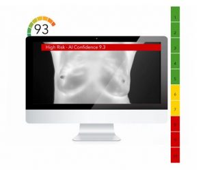
AI Talos is an AI-powered software reinventing how thermographers all around the world can detect breast cancer using deep learning and thermal imaging methods, for more information please visit our website at www.aitalos.com or have a look at our LinkedIn public page at:   https://www.linkedin.com/company/ai-talos

content
January 3, 2021
Breast Cancer Detection Using Infrared Thermal Imaging and a Deep Learning Model
January 3, 2021

One of the most common diseases that play a leading role in the death of women is breast cancer. The early and precise diagnosis of breast cancer plays a pivotal role to improve the prognosis of patients with this disease. According to many studies and extensive researches, such abnormalities can be detected and treated at primary stages of the disease making sure that highly accurate and precise techniques and soft-wares are used. Over the past 20 years, many applications have been found in the medical profession for thermal imaging such as mammography which is often used for breast cancer diagnosis. However, the potential side effects of using mammography and the rate of false positives discourages patients and physicians to use this method. In this article we focus on diagnosing breast cancer using medical imaging techniques through state-of-the-art artificial deep neural network approaches.

Previous Studies

From 2002-2010 several researchers have explored the limitations of mammography as a screening tool for detection of breast cancer. Despite the preference for mammography for the past several decades, the need for new techniques to overcome the limitations of mammography as a technique has emerged. Hence, some others focused on the neural network modalities and other innovative techniques for solving the problem of breast cancer.

Breast Cancer Detection Using Infrared Thermal Imaging

As we know, the sooner the anomaly is found, and the sooner treatment is consequently begun, and the better the chances of success. Moreover, it is important to highlight that the image processing has a powerful NIRF light signal, so that the image taken contains a lot of information that is very close to the actual state of the breast. Through the review of the articles, we found out the importance of image processing, which is currently performed well by a human being but which is not yet adequate when performed by artificial intelligence methods. This highlights the need for a Computer Assist Device (CAD) that will help us to better understand the thermal images captured by our different thermal imaging cameras. In this context a CAD will be a deep neural network with an SVM model as a classifier, as shown in Figure 10 (assuming it is already trained) that will take the thermal images in, and as output classify the images as containing cancer or not. We should clarify that the deep learning module will output the probability of a breast’s thermal image being classified as sick (having cancer) or healthy (without cancer).

Figure 10. Illustration of our model

Why using a new model?

In section 2, we outlined previous and new techniques used for the detection and prevention of breast cancer. The model that we present in this paper takes advantage of two main factors:

  • It uses a deep neural network which is modified at the last fully connected layer in such a way as to obtain a powerful binary classification (sick breast or healthy breast).
  • A second well known classifier (SVM) is coupled to that, and is involved only in the case of an uncertainty in the output of the DNN.

Conclusion

Reviewing the article, it is obvious that work in the area of breast cancer detection from a computer scientist point of view could be a valuable contribution to the field. With this in mind, we presented the techniques most commonly used to detect breast cancer, and their strengths and weaknesses. One technique in particular appeared to have a promising future, because of its non-immersive property and the significant amount of data that needs to be processed with more efficient techniques. Infrared imaging coupled with an agent previously administered to a patient can lead to a very accurate tumor detector.


References:

Mambou, S.J.; Maresova, P.; Krejcar, O.; Selamat, A.; Kuca, K. Breast Cancer Detection Using Infrared Thermal Imaging and a Deep Learning Model. Sensors 2018, 18, 2799.

FDA: Thermography is no substitute for Mammography!
January 2, 2021

FDA: “There is no valid scientific data to show that thermographic devices, when used on their own or with another diagnostic test, are an effective screening tool for any medical condition, including the early detection of breast cancer or other diseases and conditions”.

The FDA is aware that health spas, homeopathic clinics, mobile health units, and other health care facilities are using thermography inappropriately as a standalone tool for breast cancer screening or diagnosis.

The FDA has received reports that these types of facilities provide false information that can mislead patients into believing that thermography is an alternative or better option than mammography. The FDA is concerned that people will believe the misleading claims about thermography. Read below to find out more about this topic.

Breast Cancer Screening: Thermography vs. Mammogram

Since 1990s, several methods have been used for the early detection of breast cancer, such as mammography and MRI. Mammography is the standard method for early detection of breast cancer, but it has one main limitation: that is, it may produce a large number of false positives. In contrast, respective standard screening techniques are used for early detection of breast cancer such as Thermography. Thermal infrared images are used to detect lesions in breasts. Since the normal temperature range of human body is between 36.5∘C to 37.5∘C, by using this technique, thermal infrared cameras are able to capture the variation of temperature between normal and abnormal breast tissues. These Thermograms detect the temperature of the different regions of the breast. Regions with warmer temperature are more likely to contain tumors than a normal tissue. This technique has several substantial benefits:

  • It is less expensive than the mammography and magnetic resonance imaging (MRI)
  • It is non-contact, non-invasive
  • It is non-radiative
  • It is a safe diagnostic procedure, in which patients feel no pain
  • This method can be used from far way
  • It is possible to simultaneously monitor a large area of the population
  • Interpretation of thermogram’s colors is easy and fast.
  • This method only records natural radiation from the surface of the skin and there is no trace of harmful rays, so is suitable for long-term and repeating use.
  • It is a fast way to monitor and observe the dynamic changes in temperature.
  • Early detection up to 10 years before the cancer turns into a full-blown size

The tool, however, has been cleared by the FDA only as a supplement to primary diagnostic tests such as mammography and not as an substitute. Helen J. Barr, M.D., director of the Division of Mammography Quality Standards in the FDA’s Center for Devices and Radiological Health states that: “You should not rely solely on thermography for the screening or diagnosis of breast cancer.”  

The agency affirmed that individuals who substitute thermography for mammography may miss the opportunity to detect cancer at its earliest and most treatable stages. The agency wrote in its safety communications statement that: “We are committed to protecting and promoting access to safe and effective breast screening devices.” 

The United States Food and Drug Administration currently accept applications for thermographic cameras as Class I Medical Devices. These are described as devices that hold no potential risk and have the lowest level of regulatory control. Consequently, Experts highly recommend that the technique should only be used in connection with other breast cancer screening processes such as mammography. Moreover, it provides suggestions for individuals getting breast cancer screening to be aware of thermography as well as recommendations for health care providers to educate patients about the limitations of thermography.

Recommendations for People Getting Breast Cancer Screening  

  • Be aware that thermography is not a substitute for regular mammograms and should not be used in place of mammography for breast cancer screening or diagnosis.
  • Have regular mammograms according to screening guidelines or as recommended by your health care provider.
  • Follow your health care provider’s recommendations for additional steps to diagnose breast cancer such as a clinical breast exam, other breast imaging (for example, breast ultrasound or MRI), or breast tissue biopsy.

Recommendations for Health Care Providers

  • Educate patients about the limitations of thermography. For example, the high false negative and false positive rates of thermography can provide misleading information that could result in a delayed diagnosis or unnecessary medical follow up.
  • Discourage the use of thermography to diagnose or screen for breast cancer.
  • Talk to patients or caregivers about safe and effective ways to screen for breast cancer including the benefits and risks of available testing options.

Reporting Problems to the FDA

If you experience an injury or adverse event with thermography, the FDA encourages you to file a voluntary report by phone at 1-800-FDA-1088 or online at MedWatch, the FDA Safety Information and Adverse Event Reporting program. Please include the following information in your reports:

  • Device Name (Brand Name)
  • Manufacturer’s Name
  • Details of Adverse Event and Medical and/or Surgical Interventions (if applicable)

Prompt reporting of adverse events can help the FDA identify and better understand the risks related to the use of medical devices.

Additional Resources

Contact Information

If you have questions about thermography, please contact www.aitalos.com or visit our linkedIn page: https://www.linkedin.com/company/ai-talos

How Thermography works
December 28, 2020

Thermography is a breast screening technique that uses thermal infrared images for detecting breast cancer in women. It is non-invasive and painless and does not involve the compression of breasts. Although thermal imaging is not definite and surroundings can affect its results but by using thermal images irregular thermal patterns can be easily recognized by detecting increases in temperature. Thermography does not involve any radiation, instead an ultra-sensitive camera is used to create high-resolution, infrared or heat images of the breasts.


How thermography works

When tumors develop, cancer cells grow. These cells need additional blood to reproduce. So the blood flow increases for this purpose, and the skin in that area will become warmer. By using digital infrared imaging, thermography can detect subtle changes in the breast by revealing areas of heat and cold.

What to expect

Thermography test is involved of the following procedure:

  1. It should take place in a doctor’s office or a healthcare clinic
  2. The patient will stand about 6–8 feet away from the camera
  3. They will have a painless, noninvasive test that does not involve compressing the breasts
  4. The procedure will last approximately 15 minutes
  5. Doctor will look for any differences between the breasts

What thermography detects

A thermograph will show changes in body and skin temperature, which may be a sign of increased metabolic activity or blood flow in one particular area of the body. If the thermography detects any abnormalities, this may not necessarily be cancer and the person should seek further screening to confirm that a lump is present.

Benefits

Reasons why thermography is wide-accepted among the medical community is listed below:

  • Thermal imaging is non-contactand non-invasive
  • The method can be used from far way
  • It’s possible to simultaneously monitor a large area of the population
  • Interpretation of thermogram’s colors is easy and fast.
  • This method only records natural radiation from the surface of the skin and there is no trace of harmful rays, so is suitable for long-term and repeating use.
  • Finally thermography is a fast way to monitor and observe the dynamic changes in temperature.

Risks

This method is non-invasive, non-contact and does not appear to pose any physical risks to a person. In addition, it includes no compression of your breasts but the test has a high rate of false-positives and false-negatives meaning that this method is not sufficiently sensitive as a diagnostic tool. Thus, another imaging technique is likely necessary after for further investigation.

In order to get more information on Thermography screening for breast cancer detection please visit our website at www.aitalos.com. AI talos is an AI-powered screening app for detecting breast cancer using thermal infrared images. By using deep learning methods and the cutting-edge technology that we have, we’ve been able to improve detection to 95.3 percent which is 30 percent better than human operators.

How fast does breast cancer grow?
December 26, 2020

Today, one of the most common diseases that play a leading role in the death of women is breast cancer. To estimate how breast cancer can develop within a year or so is very difficult. There are different types of breast cancer that can grow at a variety of rates and several factors can have a potential impact on its growth and chance of developing.

In this article, we take a look at how quickly breast cancer might develop, types, symptoms and different ways of treating patients using AI methods.

How fast does breast cancer grow?

Since breast cancer can affect each person differently, it is not an easy task to calculate exactly how fast it can develop. Breast cancer progression and its chance of spreading depends on several factors:

  1. Types of breast cancer

Breast cancer can be invasive and non-invasive. Non-invasive breast cancer will not spread beyond the connective tissues. On the contrary, invasive breast cancer will spread to the surrounding connective tissues and other areas of the body.

  1. Grades of breast cancer (1-3)

Doctors will grade breast cancer from 1 to 3. Grade 1 is a slower-growing cancer and grade 3 is a faster-growing cancer. A higher grade means this type of cancer is more likely to grow faster and spread to other parts of the body.

  1. Stages of breast cancer (0-4)

Doctors put breast cancer progression in the category of 0-4 stages. This categorization is a substantial factor for making decisions when you are treating the patients.  

Stage 0. Non-invasive. only present in the ducts or the lobules.

Stage 1. Invasive,small and near the primary site.

Stage 2. Invasive, tumors may be larger than in stage 1, and the cancer may have spread to the lymph nodes.

Stage 3. Invasive, tumors may be larger, and cancer has spread to the lymph nodes, possibly to several. Breast cancer at this stage has not spread to other organs.

Stage 4. Breast cancer has developed in other areas of the body outside the breast and lymph nodes, often in the bones, lungs, brain, or liver.

Created for the National Cancer Institute, http://www.cancer.gov

Stage 4 cancer that has already spread to other areas of the body is more likely to spread faster.

Personal factors

American Cancer Society estimates about the 5-year survival rates for people at different stages of breast cancer. Breast cancer growth within a year will often depend on personal factors, including:

  • age at diagnosis
  • hormone status
  • family history of breast cancer
  • exposure to alcohol, cigarettes, or pollution
  • previous history of cancer

Signs and symptoms of breast cancer

According to Centers for Disease Control and Prevention (CDC), some people with breast cancer experience no symptoms at all. But people with breast cancer may experience a range of symptoms, but not all people will experience the same symptoms. Some signs and symptoms of breast cancer include:

  • a lump or mass in the breast tissue
  • pain, swelling, or redness on any part of the breast
  • dimpling of the skin covering the breast
  • unusual nipple discharge
  • flaking skin on or near the nipple
  • change in the shape or size of the breast

Detection

Several standard screening techniques are used for early detection of breast cancer such as Mammography, Thermography, Biopsy and self-examination.

Mammography: a breast cancer screening technique consisting of low-dose x-rays of the compressed breast. Clinical organizations recommend having mammograms every year beginning at age 40.

Thermography: Thermal infrared images are used to detect lesions in breasts. These Thermograms detect the temperature of the different regions of the breast. Regions with warmer temperature are more likely to contain tumors than a normal tissue.

Biopsy: This is a procedure that involves removing a sample of tissue from the breast for further examination.

Self-examination: Before women can experience any signs of cancer such as lumps or tumors, they can diagnose it with self-examination. This method can also help women to find breast diseases such as a lump, pain or nipple discharge.

Treatment

Breast cancer treatment plans depend on the type and stage of the cancer, as well as a person’s overall health status and their personal preferences. Treatments for breast cancer are either local or systemic.

Local treatment: remove cancer in a particular area of the body

Systematic treatment: destroy cancer throughout the body.

The AI Talos breast screening app provides women with access to an online breast cancer screening tech for early detection. Please visit www.aitalos.com for more information. Early detection can save your life.

FAQs about Thermography
December 19, 2020
  1. What is breast cancer Thermography?

Thermographies are thermal infrared images that are used to detect lesions in breasts. These Thermographies detect the heat pattern and blood glow of the different regions of the breast. Regions with warmer temperature are more likely to contain tumors than normal tissue. The infrared thermal imaging method can analyze breast cancer using dynamic Thermograms and also it can selectively optimize the contrast in areas of dense tissues in young women.

  1. What are the advantages of Thermography?

This techniques has respective substantial benefits:

  • It is less expensive than the mammography and magnetic resonance imaging (MRI)
  • It is non-contact, non-invasive
  • It is non-radiative
  • It is a safe diagnostic procedure, in which patients feel no pain
  • This method can be used from far way
  • It is possible to simultaneously monitor a large area of the population
  • Interpretation of Thermogram’s colors is easy and fast.
  • This method only records natural radiation from the surface of the skin and there is no trace of harmful rays, so is suitable for long-term and repeating use.
  • It is a fast way to monitor and observe the dynamic changes in temperature.
  • Early detection up to 10 years before the cancer turns into a full-blown size

  1. Is it an alternative method for Mammography?

Thermography is not an alternative method for Mammography. It is less sensitive than Mammography and can sometimes find cancerous results that are not present. Mammography has also false positives and can miss cancerous cells but experts say you should not substitute it for Mammography. The combination of two methods is likely necessary for further investigation.

  1. Who should get Thermography?

For women under the age 50 and those with dense breasts.

  1. How is the procedure going?
  • Do not wear perfume or deodorants on the day of the testing
  • You will get undressed so that your body become acclimated to the temperature of the room
  • You will stand in front of the imaging system
  • The technician takes a series of 6 images ( front and side views) of your breasts
  • The timing will be about 30 minutes
  • The doctor will analyze your images and you get the result within a few days after the test
  1. What are the possible side effects?

This method is non-invasive, non-contact and non-radiative. In addition, it includes no compression of your breasts but the test has a high rate of false-positives meaning that another imaging technique is likely necessary after this procedure for further investigation.

  1. Should I get a Thermogram or Mammogram?

Before taking a breast cancer screening test ask your doctor some questions about your high risk of getting breast cancer, the risks and benefits of getting these tests, what happens if you get false-positives and ask whether you just need Thermography or other additional breast cancer screening tests.

In order to get more information on Thermography screening for breast cancer detection please visit our website at www.aitalos.com. There you can use our top-notch AI guided software for breast cancer detection using thermal images for a thorough self-examination.

Thermography Vs. Mammography: which is the best method to detect breast cancer?
December 19, 2020

According to the researches in 2019, it has been totaled that about 30% of cancers diagnosed in women is breast cancer. Breast cancer is the most commonly cancer type detected in American women. Fortunately, breast cancer detecting techniques have been provided women with a chance to fight against this beast. But this probing question that which method is the best one to use and why is still unresolved. In this article, we will take a look at what breast cancer screening is and find the differences between two famous and popular breast screening techniques: Thermography and Mammography and find out which method is the best one to use.

Breast Cancer Screening  

Breast cancer screening is an important factor in diagnosing breast cancer at an early stage before a person can display signs and symptoms of being sick. In breast cancer screening we use different screening methods (Mammography, Thermography or biopsy) to look into a person’s body and keep track of all the anomalies detected. Hence, we can have an early and promising treatment and healing.

Different types of screening methods

Since 1960s when there was a strong tendency to use computers in order to assist physicians in identifying the abnormal areas and having a second opinion besides the doctor’s detection, breast cancer screening methods have come a very long way. For a very long time, the best standard method was Mammography. Nowadays, other screening methods such as Thermography, biopsy, Ultrasound and etc. are introduced. In this article we will have a quick look at the differences between Mammography and Thermography and the benefits and drawbacks of using these methods in detecting breast cancer.

Mammography

Mammography is a breast cancer screening technique consisting of low-dose x-rays of the compressed breasts. Clinical organizations recommend having mammograms every year beginning at age 40. Before women can experience any signs of cancer such as lumps or tumors, mammography can diagnose it with self-examination. This method can also help women to find breast diseases such as a lump, pain or nipple discharge.

Benefits of getting Mammograms:

  • Cancer detection long before display of any symptom
  • Reducing the risk of dying from breast cancer by 19% overall

Drawbacks of getting Mammograms:

  • Exposure to radiation
  • Uncovering non-cancerous changes, resulting in false-positives
  • Less accurate detection for women with dense breast tissue and breast implants
  • Uncomfortable procedure, involving of breast compression

Thermography

Thermographies are thermal infrared images that are used to detect lesions in breasts. These Thermographies detect the temperature of the different regions of the breast. Regions with warmer temperature are more likely to contain tumors than normal tissue. Although thermal imaging is not definite and surroundings can affect its results but by using thermal images irregular thermal patterns that are not visible in normal mammograms can be easily recognized.

Benefits of Thermography:

  • It is non-invasive and painless
  • It is Non-radiative
  • Capturing the entire body: detecting the heat and radiation coming from the body
  • Getting a better sense of inflammation coming from teeth ( a hidden source of breast tissue abnormalities)
  • Measuring the temperature changes
  • Early detection up to 10 years before the cancer turns into a full-blown size

Drawbacks of Thermography:

  • Another imaging technique is likely necessary after this procedure to further investigate.

Thermography can be particularly a very useful alternative tool for screening the breast cancer annually, because then you can compare the current examination with previous tests and if there is any difference then it is even a more powerful predictor. If you haven’t had your test yet, you can contact us at www.aitalos.com and there we provide you with all the necessary information about breast cancer Thermography.

Thermography: Information and Overview
December 11, 2020

Breast cancer can happen in both men and women. But it is the most prevalent type of cancers that is diagnosed among women. It can be detected as a lump in breast tissue that is different from the rest. Other signs may include: tissue thickening/ one breast becoming larger or smaller/ nipple changing position / pain in part of breast or armpit and etc.

Biopsy of the affected area of breasts is used to diagnose most types of breast cancers. Moreover, there are other types of breast cancer that need to be examined with specialized laboratory methods. Since 1990s, several methods have been used for the early detection of breast cancer, such as mammography and MRI. Mammography is the standard method for early detection of breast cancer, but it has one main limitation: that is, it may produce a large number of false positives. Here we will take a look at what Mammography is:

“ Mammography is breast cancer screening using low-dose x-rays. Medical organizations suggest having mammograms every year beginning at age 40. Before women can experience any signs of cancer, mammography can diagnose it. This method can also help women to find breast diseases such as a lump, pain or nipple discharge.”

In contrast, respective standard screening techniques are used for early detection of breast cancer such as Thermography. Thermal infrared images are used to detect lesions in breasts. These Thermograms detect the temperature of the different regions of the breast. Regions with warmer temperature are more likely to contain tumors than a normal tissue. This technique has several substantial benefits:

  • It is less expensive than the mammography and magnetic resonance imaging (MRI)
  • It is non-contact, non-invasive
  • It is non-radiative
  • It is a safe diagnostic procedure, in which patients feel no pain
  • This method can be used from far way
  • It is possible to simultaneously monitor a large area of the population
  • Interpretation of thermogram’s colors is easy and fast.
  • This method only records natural radiation from the surface of the skin and there is no trace of harmful rays, so is suitable for long-term and repeating use.
  • It is a fast way to monitor and observe the dynamic changes in temperature.
  • Early detection up to 10 years before the cancer turns into a full-blown size


Since the normal temperature range of human body is between 36.5∘C to 37.5∘C, by using this technique, thermal infrared cameras are able to capture the variation of temperature between normal and abnormal breast tissues.

AI application for breast cancer Thermography

According to several studies, by using highly well-aimed and accurate soft-wares, breast cancer can be detected and treated at early stages of the disease. Several applications have been found in the medical profession for thermal imaging. In recent years, due to advancements in image processing techniques, Thermography has been able to achieve more than other methods such as Mammography. Thermography soft-wares (CAD) serves the need of physicians for an Al-guided support for breast cancer Thermography, which is an decisive factor in validating what they speculate, detecting what they potentially did not observe throughout the process and taking notice of highly dubious studies and researches that need to be brought to more attention.

What is the difference between Thermography and Mammography?
December 10, 2020

If you are a woman, your number one risk of cancer is breast cancer and traditionally one of the most preventive screening methods is Mammography. According to one research of 50 different cancer patients, 46 cases were detected using mammograms and 44 cases were detected using thermograms. The ones that were missed using mammograms, thermograms were able to identify and the cancer cases that were missed using thermograms, mammograms could detect. Thus, many people prefer to use both of the methods in order to catch all the cases. (Dr. Troy Spearl, Synapse: Center for Health and Healing)

In this paper, we examine the differences between mammography and thermography and have a look at all the pros and cons of both methods.

Doctor and patient making a mammography

Mammography is breast cancer screening using low-dose x-rays. Medical organizations suggest having mammograms every year beginning at age 40. Before women can experience any signs of cancer, Mammography can diagnose it. This method can also help women to find breast diseases such as a lump, pain or nipple discharge. now, let’s have a look at the cons of Mammography:

The cons of Mammograms:

  • Stressful situation that patient experiences doing mammography
  • Over-diagnose of Mammograms: a lot of people go through cancer treatments even when they did not need to
  • Late identification: it would be better if we could identify breast cancer much earlier
  • More exposure to unnecessary radiation
  • More invasive

So, even though this method can serve as a screen to show breast cancer, it is not the best method to use.

When comparing the pros and cons of Mammography and Thermography, the actual cons of Thermography are only the cases that are missed and if you use ultra-sound method combined with Thermography you can check both sides (anatomical and physiological) of an image. But when it comes to the pros of Thermography, it can really check for the things are not visible in normal mammograms and also it can identify some of the certain types of inflammatory cancers even before they show up. Here, we will take a look at the pros of Thermography:

The pros of Thermography:

  • It is non-invasive and painless
  • It is Non-radiative
  • Capturing the entire body: detecting the heat and radiation coming from the body
  • Getting a better sense of inflammation coming from teeth ( a hidden source of breast tissue abnormalities)
  • Measuring the temperature changes
  • Early detection up to 10 years before the cancer turns into a full-blown size

Thermography can be particularly a very useful alternative tool for screening the breast cancer annually, because then you can compare the current examination with previous tests and if there is any difference then it is even a more powerful predictor.

This website uses cookies to improve your experience. Cookie Policy